Treatment & Management Treatment & Management

The most common indications for adults undergoing cardiac surgery are coronary artery and valve disease. These procedures are usually aimed at revascularisation or improving valve function.

While the majority of revascularisation procedures are completed percutaneously, CABG surgery is preferred for multi-vessel CAD, disease of the left main coronary artery, as well as for diffuse disease not amenable to PCI. Heart valve disease is either stenotic or regurgitant with the aortic and mitral valves most commonly affected.

For people with severe, intractable heart failure, ventricular assist devices or cardiac transplantation may be recommended.

Cardiac surgery via a median sternotomy

A median sternotomy involves the surgical division of the sternum to gain access to the heart.  This procedure is commonly used for CABG surgery and valve repairs or replacements.

A sternotomy can be full (involving a 20-25 cm incision to split the entire sternum) or partial (involving a 10-13 cm incision splitting the upper third of the sternum).

Post-operative complications may include:

Minimally invasive cardiac and percutaneous coronary interventions

Minimally invasive cardiac surgery and PCIs are less traumatic than surgery via median sternotomy. There are usually fewer complications, faster recovery, hospital stays tend to be shorter and patients are able to return to usual functional activities more quickly. 

See information on Percutaneous coronary interventions for revascularisation and valve replacement.

For more information on minimally invasive cardiac surgical procedures see http://www.hopkinsmedicine.org.

Ventricular assist devices (VADs)

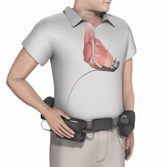
VADs augment cardiac output in patients with severe end-stage HF. They have historically been employed as a temporary ‘bridge to a cardiac transplantation’ but in some countries, a number of devices have now been approved as lifelong ‘destination therapy’. The set up for a VAD is shown in the diagram below.

Figure 1: Left ventricular assist device installation and setup

Cardiac transplantation

Cardiac transplantation is considered for those patients with severe HF who have chronic symptoms despite maximal medical therapy.

Conditions where cardiac transplantation might not be considered include:

  • Active infection
  • Cancer
  • Irreversible pulmonary hypertension
  • Severe diabetes
  • Multi-organ disease or significant co-morbid disease
  • Advanced age
  • Non-compliance with medication and care plan
  • Drug / tobacco / alcohol abuse

Respiratory function should be assessed in all patients who have undergone cardiac surgery and where possible, patients should also be assessed in the pre-operative phase. Post-operative physiotherapy is recommended to determine appropriate respiratory intervention and to facilitate enhanced ventilation and functional recovery through early mobilisation.

Positive expiratory pressure (PEP) devices improve early post-operative respiratory function and even improve early post-operative functional capacity after cardiac surgery.[#westerdahl-e-lindmark-b-eriksson-t-et-al.-2005,#stein-r-maia-cp-silveira-ad-et-al.-2009] Current evidence does not support the routine practice of preventative deep breathing exercises to reduce respiratory complications for patients after cardiac surgery.[#pasquina-p-tramler-mr-walder-b.-2003]

Approximately 30% of post-cardiac surgery patients develop musculoskeletal complications involving the shoulders, chest and upper limbs. Post-sternotomy pain may significantly interfere with comfort, function and sleep.[#stiller-k-mciness-m-huff-n-et-al.-1997,#kalso-e-mennander-s-tasmuth-t.-et-al.-2001]

Harvesting of the internal mammary artery contributes to 38.5% of patients experiencing shooting, intermittent pain of the anterior chest wall on the harvest side.[#el-ansary-d-adams-r-ghandi-a.-2000]

Tenderness may be observed on palpation of manubrium, sternocostal joints and anterior rib cage and numbness with or without allodynia of the anterior intercostal nerves T1-T2 and T5-T6.  Pain and movement dysfunction can also emanate from the graft donor site and needs to be assessed and managed as for any other wound.

When assessing pain note:

  • Onset and temporal pattern
  • Location
  • Quality/nature
  • Intensity
  • Aggravating and relieving factors
  • Previous treatment
  • Effect on physical and social function

Following an open cardiac surgical procedure, patients should be encouraged to:

  • Take pain analgesia as prescribed as it is a normal part of recovery for pain and discomfort to be present for up to 6-9 weeks following surgery.
  • Relieve pain with warm showers, heat packs and ice packs
  • Undertake shoulder, neck and back exercises to reduce chest pain and discomfort
  • Consult their medical practitioner and health care professional should any symptoms arise or persist or interfere with everyday tasks
  • 'Listen to their body' as it will set their safe limits for activity.

Wound care

Cardiac surgery patients will have one or more wounds to manage. Clinicians can positively influence the wound healing process and help prevent abnormal wound healing outcomes by fostering and delivery comprehensive wound management.

The post-operative management of the surgical wound can be straight forward.

The general principles of care are:

  • Keep the wound clean and dry
  • Assess for any complications
  • Remove sutures or staples when the wound is healed
  • Notify the clinical team immediately when signs of complications present

The specifics of wound care such as the selection of dressings and the time a dressing remains in place will be dependent on the local hospital protocol and clinicians should familiarize themselves with these and manage wound healing in accordance with local guidelines.

Note that patients are often more comfortable if the wound is covered for the first couple of weeks.

If wound complications arise, clinicians should be able to assess and identify the causal factors so that they can be managed effectively and complete wound healing can be achieved. [#oldfield-a-burton-f.-2009]

Educating the patient and their families about the importance of immediate and proper wound care should highlight the need for meticulous cleaning habits, particularly in the first 3 weeks following surgery, and risk factor modification like poor nutrition status and smoking.

Scar assessment

Regular, routine scar assessment will facilitate early identification of a problematic scar. This assessment should include both a subjective and objective assessment. The patient’s own subjective views about the scar, including pain experienced, appearance and sensitivity is important and may influence the patient’s quality of life.

The objective aspects of the scar assessment include its size, shape, colour, texture and pliability.

There are many useful objective scar assessment tools available which offer a means of obtaining quantitative measurements which are important in evaluating treatment efficacy.

Scarring represents the final stage in the normal wound healing process. Clinicians should reassure patients that it is usual to have all or some swelling, slight redness, numbness, itching and sensitivity to touch in the early post-operative months. Clinicians and their patients can help minimise these problems after cardiac surgery by following the patient tip sheet for Managing Scars after Heart Surgery.

The earlier a clinician or patient recognises the signs of abnormal scarring, the better is the likely outcome.

Patients should be advised to seek advice if the scar:

  • Is thick and raised
  • Is very sensitive and not improving
  • Changes in redness, swelling, tenderness or a discharge develops
  • Has not settled by 6 months post operatively

Factors that need to be considered prior to electing to treat a scar include:

  • Whether the scar is worsening or improving
  • The anatomical location of the scar
  • Patient’s presenting symptoms
  • Presence and/or severity of functional impairment (i.e, does the scar affect mobility)
  • Stigma associated with the scar and its impact on the patient’s quality of life
  • Likelihood of improvement with treatment

Scar management

Scars usually begin to develop 6-8 weeks post-surgery after complete re-epithelisation, and continue a process of maturation for up to 2 years. [#teot-l-roques-c-otman-s-brancati-a-et-al.-2012] While the effect of the scarring on many individuals remains non-problematic some scars may be aesthetically unappealing and restrict range of motion.

Abnormal scars are described as being:

  • Keloid
  • Hypertrophic
  • Atrophic
  • Contracted

The most common problems from scarring include: [#chen-ma-davidson-tm.-2005]

  • Pain
  • Disfigurement
  • Psychological stress
  • Loss of motion from contracture
  • Pruritus

Treatment of abnormal scarring

If a scar causes aesthetic or functional problems then treatment may be considered.

The scar treatment can be either non invasive (e.g., silicone gel sheeting, Vitamin E cream and pressure and tissue compression) or evasive (e.g., corticosteroid injection or surgical excision). [#mustoe-ta-cooter-rd-gold-mh-et-al.-2002]

Scar massage

Scar massage is used to:

  • Soften and flatten scars
  • Keep the scar tissue flexible
  • Decrease sensitivity
  • Prevent adhesions forming under the skin which can limit mobility of the chest

Massage guidance:

  • Check that the wound has healed and do not massage if any weeping or bleeding is evident
  • Use moisturisers such as vitamin E cream, sorbolene, or bio oil
  • Use the ball of the thumb in circular motions applying sufficient pressure to blanch the finger nail
  • Mobilise the tissue around the scar to break up any adhesions and avoid rubbing the surface of the scar
  • The patient should massage the area at least 3 times a day for 5-10 minutes at a time

Scar desensitisation

Often the chest area and the scar can become very sensitive. Patients can attempt to desensitise their scar by following a graded de-sensitisation program as outlined in Managing Scars after Heart Surgery. Silicone gel may also be used to help soften and sooth the scar. If symptoms persist the patient should be medically reviewed as they may require treatment with antibiotics or a topical steroid.

Exercise rehabilitation is recommended for people following cardiac surgery. See Exercise/ post surgical care for specific information about exercise considerations for these patients.

  • Westerdahl E, Lindmark B, Eriksson T, et al. Deep-breathing exercises reduce atelectasis and improve pulmonary function after coronary artery bypass surgery. Chest 2005;128:3482-3488.

    westerdahl-e-lindmark-b-eriksson-t-et-al.-2005
  • Stein R, Maia CP, Silveira AD, et al. Inspiratory muscle strength as a determinant of functional capacity early after coronary artery bypass graft surgery. Arch Phys Med Rehab 2009;90:1685-1691.

    stein-r-maia-cp-silveira-ad-et-al.-2009
  • Pasquina P, Tramler MR, Walder B. Prophylactic respiratory physiotherapy after cardiac surgery: systematic review. BMJ 2003;327:1379-1381.

    pasquina-p-tramler-mr-walder-b.-2003
  • Stiller K, McIness M, Huff N, et al. Do exercises prevent musculoskeletal complications after cardiac surgery? Physiother Theory Pract 1997;13:117-126.

    stiller-k-mciness-m-huff-n-et-al.-1997
  • Kalso E, Mennander S, Tasmuth T. et al. Chronic post-sternotomy pain. Acta Anaesthesiol Scand 2001;45:935-939.

    kalso-e-mennander-s-tasmuth-t.-et-al.-2001
  • El-Ansary D, Adams R, Ghandi A. Musculoskeletal and neurological complications following coronary artery bypass graft surgery: a comparison between saphenous vein and internal mammary artery grafting. Aust J Physiother 2000;46:19-25.

    el-ansary-d-adams-r-ghandi-a.-2000
  • Oldfield A, Burton F. Surgical Wounds: Why do they dehisce? Wound Essentials 2009; Vol 4: 84-91.

    oldfield-a-burton-f.-2009
  • Téot L, Roques C, Otman S, Brancati A, et al. Managing Scars: Measurements to Improve Scar Management. Measurements in Wound Healing. 2012. Springer London, London. 2012. p291-312.

    teot-l-roques-c-otman-s-brancati-a-et-al.-2012
  • Chen MA, Davidson TM. Scar management: prevention and treatment strategies. Curr Opin Otolaryngol Head Neck Surg 2005;13:242-247.

    chen-ma-davidson-tm.-2005